Our Office
28-02 Fair Lawn Ave
Fair Lawn, NJ 07410
Existing Patients: (201) 475-2225
New Patients: (201) 204-0157
Visit Us Online

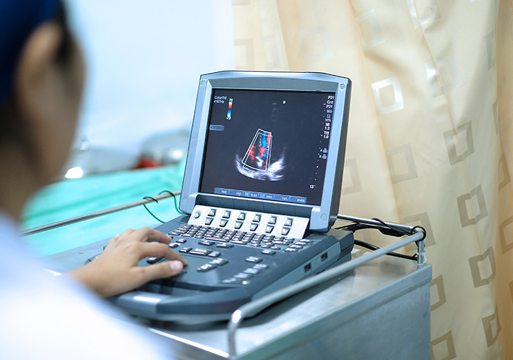
3D echocardiogram provide a cardiologist with real-time imaging of the heart’s anatomy allowing to quickly diagnose majority of cardiac problems including valvular and congenital heart conditions. Echocardiogram uses high-pitched sound waves to produce an image of the heart and can evaluate its structure, function and motion.
This evolving, leading-edge technology allows a cardiologist to clearly locate defects, tears and even clots. In addition, a 3D view provides additional ways to image the heart. This adds up to increased accuracy in diagnosing heart conditions because a cardiologist is able to view things a 2D test might miss. 3-D echocardiography allows precise ejection fraction evaluation comparable to other three-dimensional modalities such as computed tomography or cardiac MRI.
3D echocardiograms are performed in conjunction with standard 2D tests and a patient does not feel any difference in the process of 3-D images acquisition.